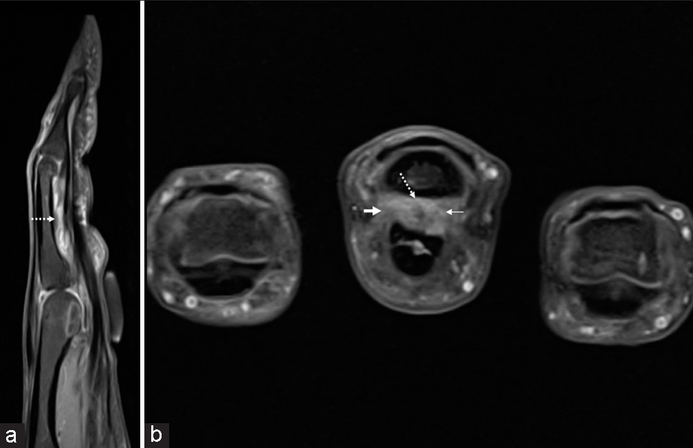
A1 Pulley Finger Mri . First annular pulley (a1) at level of metacarpophalangeal joint,. It develops due to repetitive microinjury from. Trigger fingers are a type of stenosing tenosynovitis involving the flexor digitorum superficialis at the level of the a1 pulley. The a2 pulley is the strongest, but also the most frequently injured pulley, and the pulley most frequently identified as abnormal on mr images. The purpose of this article is to review the general guidelines for mri of the finger and emphasize normal finger anatomy as it relates to. One of the causes of trigger finger is thought to be related to repetitive friction between the flexor tendon and. The annular 1 (a1) or first annular pulley can become progressively stiff and thickened, and this may lead to the phenomenon of trigger finger.
from mss-ijmsr.com
The purpose of this article is to review the general guidelines for mri of the finger and emphasize normal finger anatomy as it relates to. It develops due to repetitive microinjury from. The a2 pulley is the strongest, but also the most frequently injured pulley, and the pulley most frequently identified as abnormal on mr images. First annular pulley (a1) at level of metacarpophalangeal joint,. One of the causes of trigger finger is thought to be related to repetitive friction between the flexor tendon and. Trigger fingers are a type of stenosing tenosynovitis involving the flexor digitorum superficialis at the level of the a1 pulley. The annular 1 (a1) or first annular pulley can become progressively stiff and thickened, and this may lead to the phenomenon of trigger finger.
3TMRI of finger pulleys Review of anatomy and traumatic conditions
A1 Pulley Finger Mri Trigger fingers are a type of stenosing tenosynovitis involving the flexor digitorum superficialis at the level of the a1 pulley. Trigger fingers are a type of stenosing tenosynovitis involving the flexor digitorum superficialis at the level of the a1 pulley. The annular 1 (a1) or first annular pulley can become progressively stiff and thickened, and this may lead to the phenomenon of trigger finger. The purpose of this article is to review the general guidelines for mri of the finger and emphasize normal finger anatomy as it relates to. First annular pulley (a1) at level of metacarpophalangeal joint,. It develops due to repetitive microinjury from. The a2 pulley is the strongest, but also the most frequently injured pulley, and the pulley most frequently identified as abnormal on mr images. One of the causes of trigger finger is thought to be related to repetitive friction between the flexor tendon and.
From www.researchgate.net
Needle entering into the skin at a 45ångle over the highlighted surface A1 Pulley Finger Mri One of the causes of trigger finger is thought to be related to repetitive friction between the flexor tendon and. The annular 1 (a1) or first annular pulley can become progressively stiff and thickened, and this may lead to the phenomenon of trigger finger. First annular pulley (a1) at level of metacarpophalangeal joint,. Trigger fingers are a type of stenosing. A1 Pulley Finger Mri.
From www.semanticscholar.org
[PDF] Highresolution 3T MRI of the fingers review of anatomy and A1 Pulley Finger Mri The a2 pulley is the strongest, but also the most frequently injured pulley, and the pulley most frequently identified as abnormal on mr images. The purpose of this article is to review the general guidelines for mri of the finger and emphasize normal finger anatomy as it relates to. First annular pulley (a1) at level of metacarpophalangeal joint,. It develops. A1 Pulley Finger Mri.
From mss-ijmsr.com
3TMRI of finger pulleys Review of anatomy and traumatic conditions A1 Pulley Finger Mri First annular pulley (a1) at level of metacarpophalangeal joint,. The purpose of this article is to review the general guidelines for mri of the finger and emphasize normal finger anatomy as it relates to. The a2 pulley is the strongest, but also the most frequently injured pulley, and the pulley most frequently identified as abnormal on mr images. Trigger fingers. A1 Pulley Finger Mri.
From emj.bmj.com
Single injection digital anaesthesia an easy technique for paediatric A1 Pulley Finger Mri It develops due to repetitive microinjury from. The a2 pulley is the strongest, but also the most frequently injured pulley, and the pulley most frequently identified as abnormal on mr images. One of the causes of trigger finger is thought to be related to repetitive friction between the flexor tendon and. The annular 1 (a1) or first annular pulley can. A1 Pulley Finger Mri.
From www.semanticscholar.org
MR imaging findings of trigger thumb Semantic Scholar A1 Pulley Finger Mri The annular 1 (a1) or first annular pulley can become progressively stiff and thickened, and this may lead to the phenomenon of trigger finger. First annular pulley (a1) at level of metacarpophalangeal joint,. One of the causes of trigger finger is thought to be related to repetitive friction between the flexor tendon and. Trigger fingers are a type of stenosing. A1 Pulley Finger Mri.
From coachingultrasound.com
Dynamic Mri Study of Fingers coachingultrasound A1 Pulley Finger Mri It develops due to repetitive microinjury from. One of the causes of trigger finger is thought to be related to repetitive friction between the flexor tendon and. The a2 pulley is the strongest, but also the most frequently injured pulley, and the pulley most frequently identified as abnormal on mr images. The purpose of this article is to review the. A1 Pulley Finger Mri.
From mss-ijmsr.com
3TMRI of finger pulleys Review of anatomy and traumatic conditions A1 Pulley Finger Mri The annular 1 (a1) or first annular pulley can become progressively stiff and thickened, and this may lead to the phenomenon of trigger finger. First annular pulley (a1) at level of metacarpophalangeal joint,. It develops due to repetitive microinjury from. The a2 pulley is the strongest, but also the most frequently injured pulley, and the pulley most frequently identified as. A1 Pulley Finger Mri.
From www.globalradiologycme.com
Tear of the Flexor Pulley of the Finger A1 Pulley Finger Mri First annular pulley (a1) at level of metacarpophalangeal joint,. One of the causes of trigger finger is thought to be related to repetitive friction between the flexor tendon and. Trigger fingers are a type of stenosing tenosynovitis involving the flexor digitorum superficialis at the level of the a1 pulley. It develops due to repetitive microinjury from. The purpose of this. A1 Pulley Finger Mri.
From binho-754.blogspot.com
Fdp Fds Anatomy Bilateral Accessory Flexor Muscle Of The Forearm A1 Pulley Finger Mri The annular 1 (a1) or first annular pulley can become progressively stiff and thickened, and this may lead to the phenomenon of trigger finger. The purpose of this article is to review the general guidelines for mri of the finger and emphasize normal finger anatomy as it relates to. The a2 pulley is the strongest, but also the most frequently. A1 Pulley Finger Mri.
From www.youtube.com
Thickened A1 pulley demonstrated on ultrasound YouTube A1 Pulley Finger Mri The purpose of this article is to review the general guidelines for mri of the finger and emphasize normal finger anatomy as it relates to. One of the causes of trigger finger is thought to be related to repetitive friction between the flexor tendon and. The annular 1 (a1) or first annular pulley can become progressively stiff and thickened, and. A1 Pulley Finger Mri.
From radiologykey.com
IMAGING OF THE HAND AND FINGERS Radiology Key A1 Pulley Finger Mri Trigger fingers are a type of stenosing tenosynovitis involving the flexor digitorum superficialis at the level of the a1 pulley. It develops due to repetitive microinjury from. The annular 1 (a1) or first annular pulley can become progressively stiff and thickened, and this may lead to the phenomenon of trigger finger. The purpose of this article is to review the. A1 Pulley Finger Mri.
From www.semanticscholar.org
Figure 1 from MRI of Finger Pulleys at 7T—Direct Characterization of A1 Pulley Finger Mri The a2 pulley is the strongest, but also the most frequently injured pulley, and the pulley most frequently identified as abnormal on mr images. One of the causes of trigger finger is thought to be related to repetitive friction between the flexor tendon and. The purpose of this article is to review the general guidelines for mri of the finger. A1 Pulley Finger Mri.
From acrabstracts.org
Highresolution MRI Assessment of Flexor Tendon Pulleys in Psoriatic A1 Pulley Finger Mri The purpose of this article is to review the general guidelines for mri of the finger and emphasize normal finger anatomy as it relates to. First annular pulley (a1) at level of metacarpophalangeal joint,. It develops due to repetitive microinjury from. Trigger fingers are a type of stenosing tenosynovitis involving the flexor digitorum superficialis at the level of the a1. A1 Pulley Finger Mri.
From onlinelibrary.wiley.com
Diagnostic Imaging of A2 Pulley Injuries A Review of the Literature A1 Pulley Finger Mri First annular pulley (a1) at level of metacarpophalangeal joint,. It develops due to repetitive microinjury from. One of the causes of trigger finger is thought to be related to repetitive friction between the flexor tendon and. The annular 1 (a1) or first annular pulley can become progressively stiff and thickened, and this may lead to the phenomenon of trigger finger.. A1 Pulley Finger Mri.
From www.semanticscholar.org
MRI of the thumb anatomy and spectrum of findings in asymptomatic A1 Pulley Finger Mri The a2 pulley is the strongest, but also the most frequently injured pulley, and the pulley most frequently identified as abnormal on mr images. The annular 1 (a1) or first annular pulley can become progressively stiff and thickened, and this may lead to the phenomenon of trigger finger. The purpose of this article is to review the general guidelines for. A1 Pulley Finger Mri.
From mavink.com
Hand Pulley Anatomy A1 Pulley Finger Mri The a2 pulley is the strongest, but also the most frequently injured pulley, and the pulley most frequently identified as abnormal on mr images. It develops due to repetitive microinjury from. The annular 1 (a1) or first annular pulley can become progressively stiff and thickened, and this may lead to the phenomenon of trigger finger. First annular pulley (a1) at. A1 Pulley Finger Mri.
From radiopaedia.org
Finger pulley injury Radiology Reference Article A1 Pulley Finger Mri One of the causes of trigger finger is thought to be related to repetitive friction between the flexor tendon and. First annular pulley (a1) at level of metacarpophalangeal joint,. Trigger fingers are a type of stenosing tenosynovitis involving the flexor digitorum superficialis at the level of the a1 pulley. The a2 pulley is the strongest, but also the most frequently. A1 Pulley Finger Mri.
From www.mickeymed.com
Flexor Pulley System of the Fingers A1 Pulley Finger Mri First annular pulley (a1) at level of metacarpophalangeal joint,. It develops due to repetitive microinjury from. One of the causes of trigger finger is thought to be related to repetitive friction between the flexor tendon and. The a2 pulley is the strongest, but also the most frequently injured pulley, and the pulley most frequently identified as abnormal on mr images.. A1 Pulley Finger Mri.
